Read the neck veins.
The JVP is one of the highest-yield signs in heart failure, and one of the most butchered. Nearly every miss is technique: bad positioning, or calling the carotid a vein.
Reviewed August 2026 · verify against current guidelines
Read the neck veins.
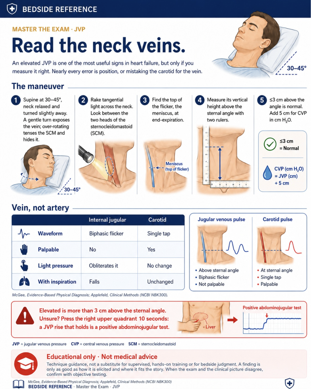
An elevated JVP is one of the most useful signs in heart failure, but only if you measure it right. Nearly every error is position, or mistaking the carotid for the vein.
The maneuver
- Supine at 30–45°, neck relaxed and turned slightly away. A gentle turn exposes the vein; over-rotating tenses the SCM and hides it.
- Rake tangential light across the neck. Look between the two heads of the sternocleidomastoid.
- Find the top of the flicker, the meniscus, at end-expiration.
- Measure its vertical height above the sternal angle with two rulers.
- ≤3 cm above the angle is normal. Add 5 cm for CVP in cm H2O.
Vein, not artery
| Internal jugular | Carotid | |
|---|---|---|
| Waveform | Biphasic flicker | Single tap |
| Palpable | No | Yes |
| Light pressure | Obliterates it | No change |
| With inspiration | Falls | Unchanged |
McGee, Evidence-Based Physical Diagnosis; Applefeld, Clinical Methods (NCBI NBK300). Technique guidance, not a substitute for supervised, hands-on training. When the exam and the clinical picture disagree, confirm with objective testing.
Sources
Verify against current guidelines and local protocol before acting.
- McGee, Evidence-Based Physical Diagnosis · Applefeld, Clinical Methods (NCBI NBK300)
Downloads
Every card for this topic — carousels and tables, print-ready for the wards or for sharing.