The falsely normal SpO₂.
The oximeter shines two wavelengths and assumes two pigments. Carbon monoxide reads as saturated; methemoglobin pins the display near 85%. Ask for co-oximetry — a routine gas calculates saturation, it doesn't measure it.
Reviewed August 2026 · verify against current guidelines
The falsely normal SpO₂.
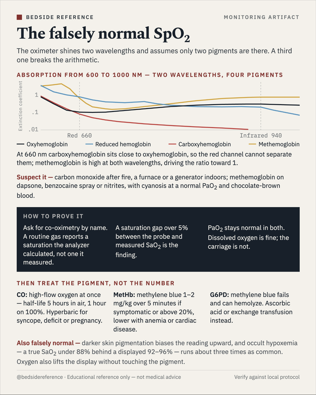
The oximeter shines two wavelengths and assumes only two pigments are there. A third one breaks the arithmetic. At 660 nm carboxyhemoglobin sits close to oxyhemoglobin, so the red channel cannot separate them; methemoglobin is high at both wavelengths, driving the ratio toward 1 — which pins the display near 85%.
Suspect it — carbon monoxide after fire, a furnace or a generator indoors; methemoglobin on dapsone, benzocaine spray or nitrites, with cyanosis at a normal PaO2 and chocolate-brown blood.
How to prove it
- Ask for co-oximetry by name. A routine gas reports a saturation the analyzer calculated, not one it measured.
- A saturation gap over 5% between the probe and measured SaO2 is the finding.
- PaO2 stays normal in both. Dissolved oxygen is fine; the carriage is not.
Then treat the pigment, not the number
| Pigment | Treatment |
|---|---|
| CO | High-flow oxygen at once — half-life 5 hours in air, 1 hour on 100%. Hyperbaric for syncope, deficit or pregnancy. |
| MetHb | Methylene blue 1–2 mg/kg over 5 minutes if symptomatic or above 20%, lower with anemia or cardiac disease. |
| G6PD | Methylene blue fails and can hemolyze. Ascorbic acid or exchange transfusion instead. |
Barker, Anesthesiology 1987 (dyshemoglobins and pulse oximetry); Sjoding, NEJM 2020 (occult hypoxemia by skin pigmentation). Educational reference only — verify against local protocol.
Sources
Verify against current guidelines and local protocol before acting.
- Barker, Anesthesiology 1987 · Sjoding, NEJM 2020
Downloads
Every card for this topic — carousels and tables, print-ready for the wards or for sharing.