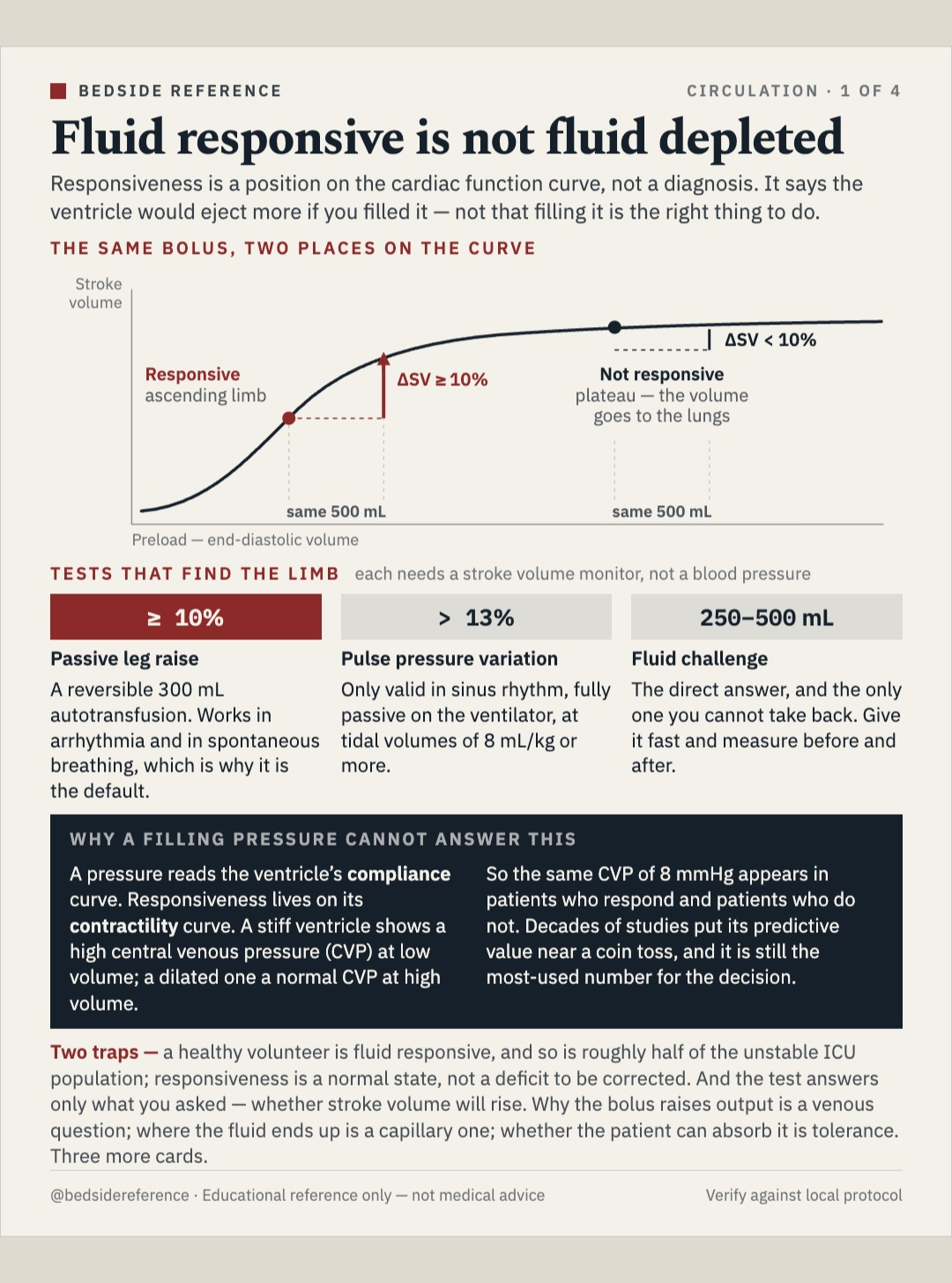
Fluid responsive is not fluid depleted.
Four cards on one decision. Responsiveness is a position on a curve, not a diagnosis. The bolus works by widening a venous gradient, most of it leaves the capillary within hours, and a responsive patient can still be intolerant. Tolerance is the permission, and it's asked first.
Reviewed September 2026 · verify against current guidelines
Fluid responsive is not fluid depleted.
Responsiveness is a position on the cardiac function curve, not a diagnosis. It says the ventricle would eject more if you filled it — not that filling it is the right thing to do.
The same bolus, two places on the curve
On the ascending limb the same 500 mL raises stroke volume by 10% or more — responsive. On the plateau the same 500 mL raises it by less than 10% — not responsive, and the volume goes to the lungs.
Tests that find the limb — each needs a stroke volume monitor, not a blood pressure
| Test | Threshold | Notes |
|---|---|---|
| Passive leg raise | ΔSV ≥ 10% | A reversible 300 mL autotransfusion. Works in arrhythmia and in spontaneous breathing, which is why it is the default. |
| Pulse pressure variation | > 13% | Only valid in sinus rhythm, fully passive on the ventilator, at tidal volumes of 8 mL/kg or more. |
| Fluid challenge | 250–500 mL | The direct answer, and the only one you cannot take back. Give it fast and measure before and after. |
The gradient, not the pressure.
Blood returns to the heart down a pressure difference. A bolus works only if it raises the upstream pressure more than the downstream one — and the heart decides which.
The equation
VR = (MSFP − CVP) / Rv. Venous return runs on mean systemic filling pressure — 8–10 mmHg in a stopped circulation — minus the central venous pressure it drains into, over venous resistance.
The same bolus, two hearts — a bolus shifts the return line right; the heart decides the rest
| Steep curve | The bolus buys flow. Flow rises sharply; CVP barely moves. |
|---|---|
| Flat curve | The bolus buys pressure. CVP rises sharply; flow barely moves. |
Two volumes, only one of which does anything
About 30% of blood volume is arterial; the veins hold about 70%, most of it unstressed. Unstressed volume fills the vessel without stretching it — it generates no pressure and adds nothing to return. Stressed volume alone sets MSFP. A pressor converts unstressed into stressed.
Three consequences that change what you do
- A rising CVP is the failure signal. If the bolus moved the pressure and not the flow, you narrowed the gradient and added congestion for nothing.
- A vasopressor is a bolus you can take back. Norepinephrine squeezes unstressed into stressed volume, raising MSFP without adding a drop.
- CVP is what the organs drain against. Kidney and liver perfusion depend on the gradient across them, so a high CVP injures from behind.
Where the fluid goes.
The classical model had fluid leaving the capillary at one end and returning at the other. It does not. Filtration runs the whole length, the lymphatics are the only way back, and the barrier is a gel you can strip off.
The revised Starling equation
Jv = LpA [ (Pc − Pi) − σ(πp − πsg) ]. One term changed and the whole model with it: the oncotic pull is set by the subglycocalyx space, not the interstitium.
Where the barrier actually sits
Glycocalyx, then the subglycocalyx space, then endothelium. Filtration continues the whole length of the capillary — it never reverses, and there is no venous reabsorption.
What the revision costs you at the bedside
| ~15% | Roughly the share of a crystalloid bolus still intravascular within a few hours of giving it in inflammation. |
|---|---|
| σ falls | Sepsis sheds the glycocalyx, so the reflection coefficient drops and the colloid advantage largely goes with it. |
| Lymph is the exit | Flow can rise several-fold, but not indefinitely. Edema is filtration outrunning that ceiling. |
Responsive, but not tolerant.
Responsiveness asks whether the output will rise. Tolerance asks what the volume will cost the lung, the kidney and the gut. They are independent, and only tolerance is a permission.
Reading tolerance — congestion upstream of each organ, not a single number
| Lung | B-lines in multiple zones, falling oxygenation, more oxygen needed during the bolus itself. |
|---|---|
| Kidney and gut | Venous congestion on Doppler, a dilated inferior vena cava that does not vary, rising abdominal pressure. |
| Right heart | A dilated right ventricle turns each further bolus into septal shift and less left-sided filling. |
Two questions, four patients
| Tolerant | Intolerant | |
|---|---|---|
| Responsive | Give it. The only quadrant where a bolus is the answer. Small aliquot, measure, repeat while the flow rises. | Buy the flow another way. The output would rise, but congestion already costs more than the flow is worth. Pressor or inotrope instead. |
| Not responsive | No benefit to buy. The volume is absorbed without harm and without gain. Look elsewhere for the cause of the shock. | De-resuscitate. Nothing to gain, harm accruing. Stop maintenance fluids, then diuretic or ultrafiltration. Most late ICU patients are here. |
Benefit falls, harm rises
- The first hours buy the most. The gradient is genuinely low and the barrier largely intact, so volume does what you intend.
- By day two it inverts. The same bolus mostly buys congestion, and a cumulative positive balance tracks with worse outcomes.
Caveats exist; this is a general framework. Educational reference only — verify against local protocol.
Sources
Verify against current guidelines and local protocol before acting.
- Monnet X, Teboul JL. Passive leg raising: five rules, not a drop of fluid! Crit Care 2015;19:18.
- Marik PE, Baram M, Vahid B. Does central venous pressure predict fluid responsiveness? A systematic review of the literature and the tale of seven mares. Chest 2008;134:172–178.
- Michard F, Teboul JL. Predicting fluid responsiveness in ICU patients: a critical analysis of the evidence. Chest 2002 (pulse pressure variation).
- Guyton AC. Determination of cardiac output by equating venous return curves with cardiac response curves. Physiol Rev 1955.
- Woodcock TE, Woodcock TM. Revised Starling equation and the glycocalyx model of transvascular fluid exchange. Br J Anaesth 2012;108:384–394.
- Malbrain MLNG, et al. Principles of fluid management and stewardship in septic shock: the four D's and the four phases. Ann Intensive Care 2018.
- Boyd JH, et al. Fluid resuscitation in septic shock: a positive fluid balance and elevated CVP are associated with increased mortality. Crit Care Med 2011.
Downloads
Every card for this topic — carousels and tables, print-ready for the wards or for sharing.