Reading a chest x-ray.
A simple, repeatable system so you read every film the same way: confirm identity, check quality (RIPE), then airway-to-soft-tissue in the same order — plus the common misses behind the diaphragm and heart.
Reviewed June 2026 · verify against current guidelines
Before you read
Confirm, then be systematic.
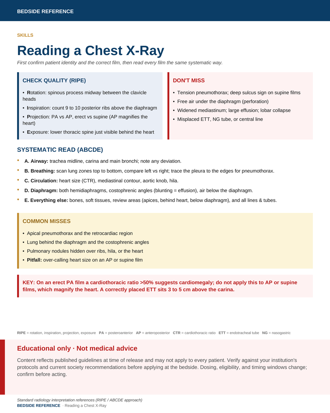
First confirm patient identity and the correct film, then read every film the same systematic way.
Check quality (RIPE)
- Rotation: spinous process midway between the clavicle heads.
- Inspiration: count 9 to 10 posterior ribs above the diaphragm.
- Projection: PA vs AP, erect vs supine (AP magnifies the heart).
- Exposure: lower thoracic spine just visible behind the heart.
Don't miss
- Tension pneumothorax; deep sulcus sign on supine films.
- Free air under the diaphragm (perforation).
- Widened mediastinum; large effusion; lobar collapse.
- Misplaced ETT, NG tube, or central line.
Systematic read
ABCDE — every film, the same way.
- A. Airway: trachea midline, carina and main bronchi; note any deviation.
- B. Breathing: scan lung zones top to bottom, compare left vs right; trace the pleura to the edges for pneumothorax.
- C. Circulation: heart size (CTR), mediastinal contour, aortic knob, hila.
- D. Diaphragm: both hemidiaphragms, costophrenic angles (blunting = effusion), air below the diaphragm.
- E. Everything else: bones, soft tissues, review areas (apices, behind heart, below diaphragm), and all lines & tubes.
Common misses
- Apical pneumothorax and the retrocardiac region.
- Lung behind the diaphragm and the costophrenic angles.
- Pulmonary nodules hidden over ribs, hila, or the heart.
- Pitfall: over-calling heart size on an AP or supine film.
On an erect PA film a cardiothoracic ratio >50% suggests cardiomegaly; do not apply this to AP or supine films, which magnify the heart. A correctly placed ETT sits 3 to 5 cm above the carina.
RIPE rotation, inspiration, projection, exposurePA posteroanteriorAP anteroposteriorCTR cardiothoracic ratioETT endotracheal tubeNG nasogastric
Sources
Verify against current guidelines and local protocol before acting.
- Standard radiology interpretation references (RIPE / ABCDE approach)
Downloads
Every card for this topic — carousels and tables, print-ready for the wards or for sharing.