The end-tidal CO₂ gap.
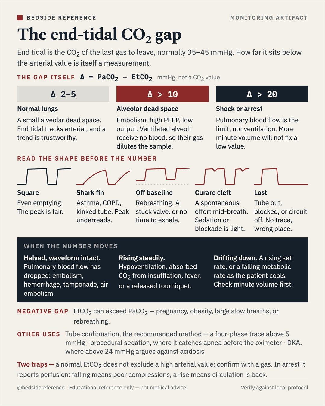
End tidal is the CO₂ of the last gas to leave, normally 35–45 mmHg. How far it sits below the arterial value is itself a measurement — and the waveform shape has to be read before the number.
Reviewed September 2026 · verify against current guidelines
The end-tidal CO₂ gap.
End tidal is the CO2 of the last gas to leave, normally 35–45 mmHg. How far it sits below the arterial value is itself a measurement.
The gap itself
Δ = PaCO2 − EtCO2 — in mmHg, not a CO2 value.
| Gap | Meaning | What it says |
|---|---|---|
| Δ 2–5 | Normal lungs | A small alveolar dead space. End tidal tracks arterial, and a trend is trustworthy. |
| Δ > 10 | Alveolar dead space | Embolism, high PEEP, low output. Ventilated alveoli receive no blood, so their gas dilutes the sample. |
| Δ > 20 | Shock or arrest | Pulmonary blood flow is the limit, not ventilation. More minute volume will not fix a low value. |
Read the shape before the number
| Waveform | What it means |
|---|---|
| Square | Even emptying. The peak is fair. |
| Shark fin | Asthma, COPD, kinked tube. Peak underreads. |
| Off baseline | Rebreathing. A stuck valve, or no time to exhale. |
| Curare cleft | A spontaneous effort mid-breath. Sedation or blockade is light. |
| Lost | Tube out, blocked, or circuit off. No trace, wrong place. |
When the number moves
- Halved, waveform intact. Pulmonary blood flow has dropped: embolism, hemorrhage, tamponade, air embolism.
- Rising steadily. Hypoventilation, absorbed CO2 from insufflation, fever, or a released tourniquet.
- Drifting down. A rising set rate, or a falling metabolic rate as the patient cools. Check minute volume first.
Negative gap
EtCO2 can exceed PaCO2 — pregnancy, obesity, large slow breaths, or rebreathing.
Other uses
Tube confirmation, the recommended method — a four-phase trace above 5 mmHg · procedural sedation, where it catches apnea before the oximeter · DKA, where above 24 mmHg argues against acidosis.
Educational reference only — verify against local protocol.
Sources
Verify against current guidelines and local protocol before acting.
- Panchal AR, et al. 2020 AHA Guidelines for CPR and Emergency Cardiovascular Care: Adult Basic and Advanced Life Support. Circulation 2020 (EtCO₂ for tube confirmation and CPR quality).
- Nagler J, Krauss B. Capnography: a valuable tool for airway management. Emerg Med Clin North Am 2008.
- Kodali BS. Capnography outside the operating rooms. Anesthesiology 2013.
- Soleimanpour H, et al. Capnography in the emergency department. Emerg Med 2012 (EtCO₂ and DKA screening).
Downloads
Every card for this topic — carousels and tables, print-ready for the wards or for sharing.