ARDS: the injured lung is small, not stiff.
The injured lung isn't stiff — it's small. A full breath lands in a lung the size of a child's, and that strain is what kills. Five cards: what counts as ARDS, the settings that protect it, the rescue ladder, what it costs the right heart, and how to get back off.
Reviewed September 2026 · verify against current guidelines
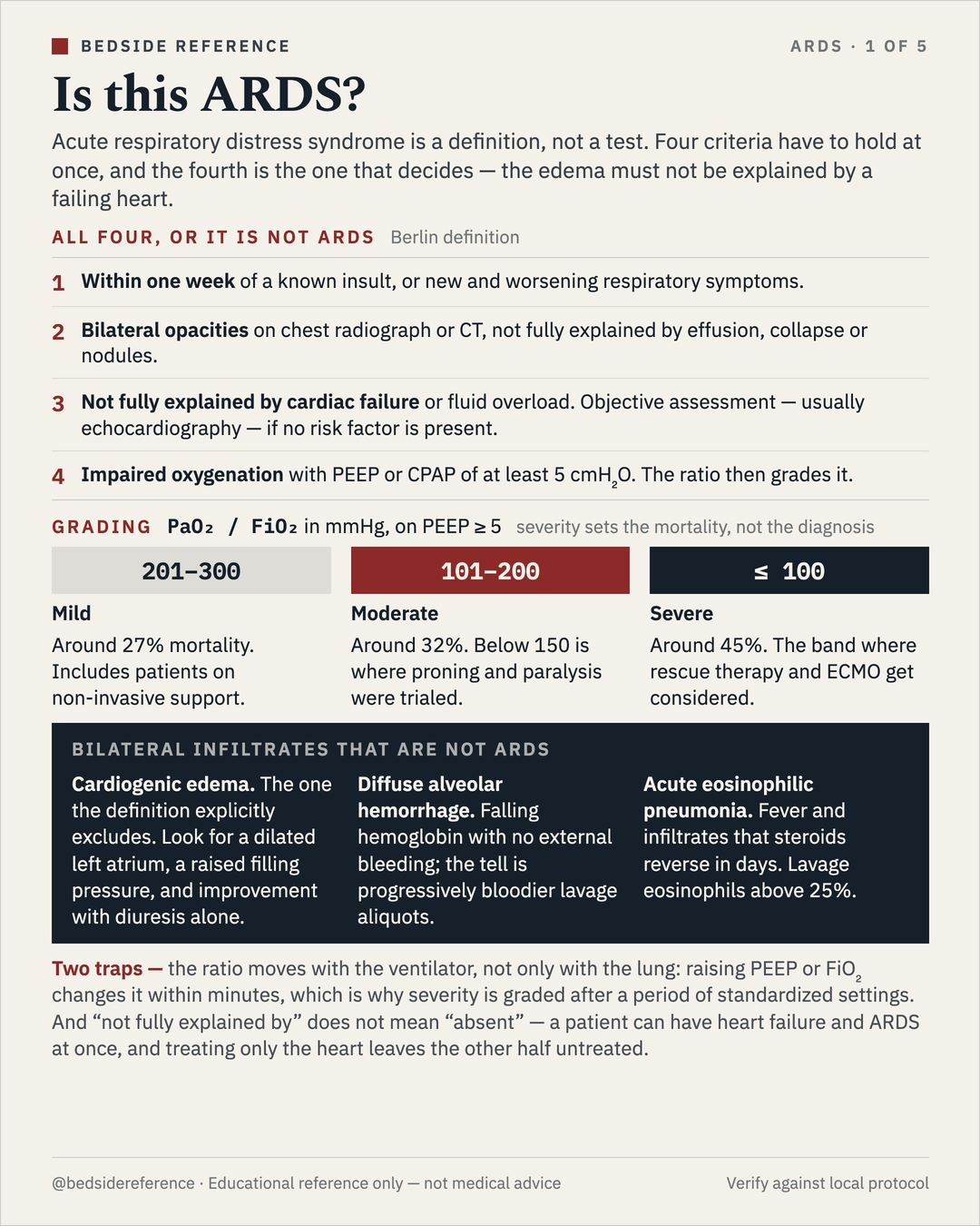
Is this ARDS?
Acute respiratory distress syndrome is a definition, not a test. Four criteria have to hold at once, and the fourth is the one that decides — the edema must not be explained by a failing heart.
All four, or it is not ARDS — Berlin definition
- Within one week of a known insult, or new and worsening respiratory symptoms.
- Bilateral opacities on chest radiograph or CT, not fully explained by effusion, collapse or nodules.
- Not fully explained by cardiac failure or fluid overload. Objective assessment — usually echocardiography — if no risk factor is present.
- Impaired oxygenation with PEEP or CPAP of at least 5 cmH2O. The ratio then grades it.
Grading — PaO₂ / FiO₂ in mmHg, on PEEP ≥ 5; severity sets the mortality, not the diagnosis
| P/F | Grade | Mortality |
|---|---|---|
| 201–300 | Mild | Around 27%. Includes patients on non-invasive support. |
| 101–200 | Moderate | Around 32%. Below 150 is where proning and paralysis were trialed. |
| ≤ 100 | Severe | Around 45%. The band where rescue therapy and ECMO get considered. |
Bilateral infiltrates that are not ARDS
- Cardiogenic edema. The one the definition explicitly excludes. Look for a dilated left atrium, a raised filling pressure, and improvement with diuresis alone.
- Diffuse alveolar hemorrhage. Falling hemoglobin with no external bleeding; the tell is progressively bloodier lavage aliquots.
- Acute eosinophilic pneumonia. Fever and infiltrates that steroids reverse in days. Lavage eosinophils above 25%.
The settings that keep the lung.
The injured lung is small, not stiff. Only a fraction of it is open to ventilate, so a normal tidal volume is delivered into a baby-sized lung — and the harm is the strain, not the volume.
Driving pressure
ΔP = Vt / Crs = Pplat − PEEP. Driving pressure is tidal volume normalized to the lung you actually have. It tracks mortality better than either term alone.
Set it in this order — each step has a number that tells you to stop
| 1 · Predicted body weight | From height and sex, never actual weight. A 1.6 m woman gets the same lung as a 1.6 m woman twice her mass. |
|---|---|
| 2 · Tidal volume 6 mL/kg | Start at 6, range 4–8. ARMA cut mortality from 40% to 31% against 12 mL/kg. |
| 3 · Plateau ≤ 30 cmH2O | Measured on a 2-second inspiratory hold. Over 30, drop Vt by 1 mL/kg at a time, to a floor of 4. |
| 4 · Driving pressure ≤ 15 | If ΔP stays high at a legal plateau, the lung is smaller than the numbers suggest. |
| 5 · PEEP from the table | Paired to FiO2, targeting PaO2 55–80 mmHg or SpO2 88–95%. |
The two PEEP tables — pick one and stay on it; the higher table suits moderate to severe disease
| FiO₂ | .30 | .40 | .50 | .60 | .70 | .80 | .90 | 1.0 |
|---|---|---|---|---|---|---|---|---|
| Lower | 5 | 5–8 | 8–10 | 10 | 10–14 | 14 | 14–18 | 18–24 |
| Higher | 5–14 | 14–16 | 16–20 | 20 | 20 | 20–22 | 22 | 22–24 |
What you accept to keep the settings
- Permissive hypercapnia. A rising CO2 is the price of a small tidal volume. Tolerate a pH down to about 7.20 before you trade lung protection for it; raise the rate first, up to 35.
- Oxygenation that looks wrong. A saturation of 88–95% is the target, not a failure to correct. Chasing a normal number with FiO2 and volume is how the ventilator does the injuring.
When oxygenation fails anyway.
Two of these save lives, one does not, and one buys time. The order is the order of the evidence.
Escalate in this order — P/F < 150 triggers the first two
| Intervention | Evidence | Verdict |
|---|---|---|
| Prone positioning | At least 16 consecutive hours. PROSEVA halved 28-day mortality, 33% to 16%. | Do it first |
| Neuromuscular blockade | ACURASYS found benefit at 48 hours; ROSE, with lighter sedation in controls, found none. | If needed |
| Inhaled vasodilator | Nitric oxide improves the P/F ratio without improving survival. A bridge, not a treatment. | Buys time |
| Recruitment maneuver | ART found higher 28-day mortality with stepwise recruitment, 55% against 49%. | Harm signal |
Why turning the patient over works
Supine, the ventral lung is overdistended while the dorsal lung collapses with its perfusion preserved — ventilation and blood flow are mismatched. Prone, the dorsal lung recruits and inflation becomes more uniform, so ventilation reaches well-perfused dorsal lung.
The right heart pays for it.
Every lever that protects the lung raises the load on the right ventricle. Acute cor pulmonale appears in roughly a fifth of moderate to severe ARDS, and the ventilator is part of the cause.
How lung protection becomes right heart failure
High PEEP, hypercapnia and hypoxic vasoconstriction → pulmonary vascular resistance rises → right ventricular afterload rises → the RV dilates and the septum shifts left → LV filling falls, and so does the output → a protected lung and a failing pump.
Four risk factors — none present, about 4% have cor pulmonale; all four, about three quarters
| Threshold | Factor |
|---|---|
| ≥ 18 | Driving pressure, in cmH2O. The one you can change tonight. |
| ≥ 48 | PaCO2, in mmHg. Permissive hypercapnia has a cost, and this is it. |
| < 150 | The P/F ratio. The same threshold that triggers proning. |
| Pneumonia | As the cause of the ARDS. Fixed, but it tells you to go looking. |
When the lung cannot be protected at all — the EOLIA thresholds
| P/F < 50 | for more than 3 hours |
|---|---|
| P/F < 80 | for more than 6 hours |
| pH < 7.25 | with PaCO2 ≥ 60 for more than 6 hours |
Getting back off.
Liberation is not a single decision at the end. It is five things you start on day one and undo in order — and the longest delays are sedation and accumulated fluid, not the lung.
Undo it in this order — each step is checked daily, not once
| 1 · Lighten the sedation | Target RASS 0 to −2 with a daily interruption. Deep sedation on day one predicts longer ventilation and higher mortality. |
|---|---|
| 2 · Settle the dyssynchrony | Watch the flow and pressure traces, not the number. Double triggering and reverse triggering are the two that injure. |
| 3 · Take the fluid back | Once the shock has resolved. FACTT bought 2.5 more ventilator-free days with a conservative strategy, without costing survival. |
| 4 · Screen, then trial | A spontaneous breathing trial of 30 to 120 minutes, on pressure support or a T-piece, once the screen below is passed. |
| 5 · Extubate, then support | High-flow or non-invasive support straight after, in the patients at risk of failing. |
The screen before the trial — a general guide; individual patients vary
| Threshold | Criterion |
|---|---|
| ≤ 0.40 | FiO2, with PEEP of 8 or less and adequate oxygenation. |
| < 105 | Rapid shallow breathing index — rate over tidal volume in liters. |
| Awake | Following commands, with an effective cough and a manageable secretion load. |
| Off pressors | Or on a low, stable dose. Shock is the commonest reason the trial fails. |
Educational reference only — verify against local protocol.
Sources
Verify against current guidelines and local protocol before acting.
- ARDS Definition Task Force. Acute Respiratory Distress Syndrome: The Berlin Definition. JAMA 2012;307:2526–2533.
- ARDS Network (ARMA). Ventilation with lower tidal volumes as compared with traditional tidal volumes for ALI and ARDS. NEJM 2000;342:1301–1308.
- Amato MBP, et al. Driving pressure and survival in the acute respiratory distress syndrome. NEJM 2015;372:747–755.
- Guérin C, et al. (PROSEVA). Prone positioning in severe acute respiratory distress syndrome. NEJM 2013;368:2159–2168.
- Papazian L, et al. (ACURASYS). Neuromuscular blockers in early ARDS. NEJM 2010;363:1107–1116.
- National Heart, Lung, and Blood Institute PETAL Network (ROSE). Early neuromuscular blockade in ARDS. NEJM 2019;380:1997–2008.
- Writing Group for the ART Investigators. Effect of lung recruitment and titrated PEEP vs low PEEP on mortality in ARDS. JAMA 2017;318:1335–1345.
- Combes A, et al. (EOLIA). ECMO for severe acute respiratory distress syndrome. NEJM 2018;378:1965–1975.
- Mekontso Dessap A, et al. Acute cor pulmonale in ARDS: prevalence, predictors, and clinical impact. Intensive Care Med 2016;42:862–870.
- NHLBI ARDS Clinical Trials Network (FACTT). Comparison of two fluid-management strategies in acute lung injury. NEJM 2006;354:2564–2575.
- Grasselli G, et al. ESICM guidelines on ARDS: definition, phenotyping and respiratory support strategies. Intensive Care Med 2023.
Downloads
Every card for this topic — carousels and tables, print-ready for the wards or for sharing.